Investigating the Microscale Mechanics of Actomyosin Networks
Author:
Bernard Iringire Nkusi ’27Co-Authors:
Faculty Mentor(s):
Bekele Gurmessa, department of Physics & AstronomyFunding Source:
Program for Undergraduate ResearchAbstract
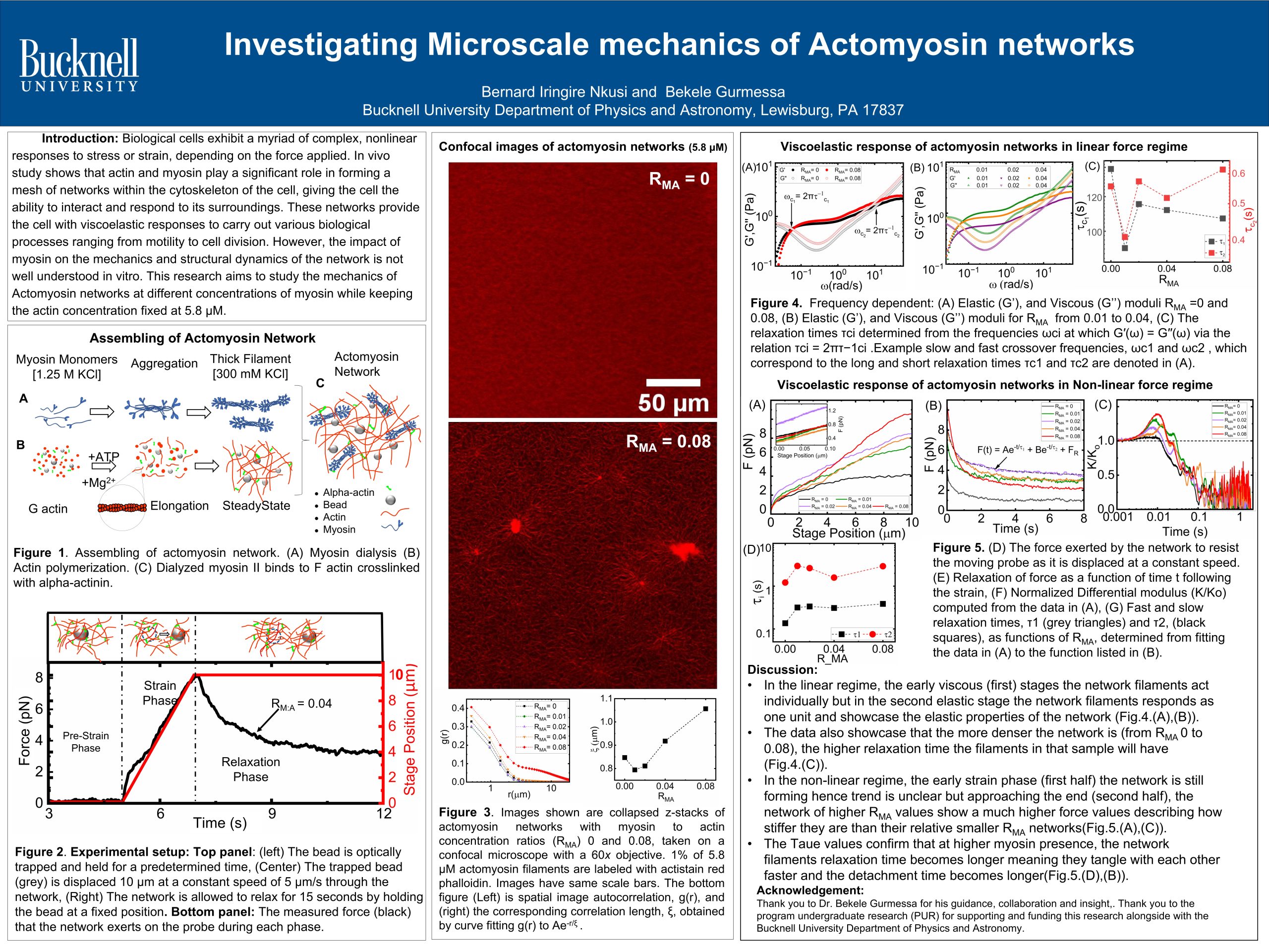
Biological cells exhibit a myriad of complex nonlinear responses to stress or strain, namely stress stiffening, softening, elastic recovery, and plastic deformation depending on the nature of the stress applied. Actin, a vital cytoskeleton protein, plays a key role in maintaining cellular stability, enabling motion, facilitating replication, and powering muscle contraction. Its filaments are entangled and crosslinked, which gives the cytoskeleton its viscoelastic response and modulates various mechanically-driven processes regulated by actin-binding proteins such as myosin. This important molecular protein-Myosin, generates contractile forces by exerting tension on actin filaments in opposing directions which creates elongated tail-to-tail thick filaments through ATP hydrolysis. The process generates forces at the piconewton level. The forces generated are crucial for orchestrating localized pulling forces during fundamental cellular processes such as division, migration, and muscle contraction. Despite the growing evidence entailing myosin-II as a key part in such microscale mechanics, the mechanical behavior/response of the network crosslinked by myosin-II is poorly understood. Beyond its role in introducing network contractility, the precise threshold concentration of myosin-to-actin and ATP ratio, to which myosin acts as a crosslinker, remains elusive.
Our project aimed to develop a series of in vitro reconstituted actomyosin networks by tuning the myosin-to-actin ratios to examine the mechanical properties and structural reorganization of these networks. This approach discerned the chemical environments conducive to actomyosin network formation, provided insight into how different ratios of myosin-to-actin affects the network’s structure and mechanical properties, and determined how the relaxation time scale depends on the concentration of myosin.