Low-Frequency, Low-Intensity Ultrasound Increases Pro-Angiogenic Protein Secretion in 3D Macrophage-Endothelial Co-Cultures
Author:
Isaac Horst ’27Co-Authors:
Faculty Mentor(s):
Dr. Olivia Boerman, Biomedical EngineeringFunding Source:
PURAbstract
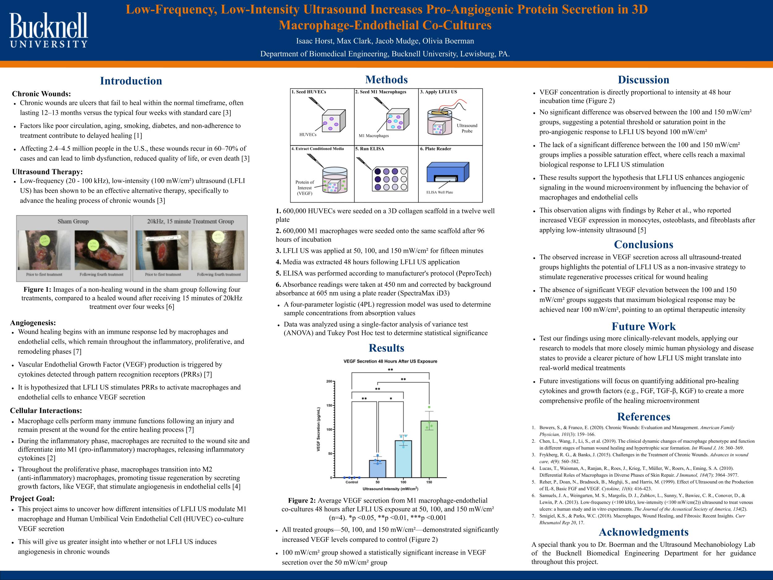
Chronic wounds often stall in the inflammatory phase, creating a significant clinical burden. This study investigated Low-Frequency, Low-Intensity Ultrasound (LFLI US) as a non-invasive intervention to trigger the proliferative phase of healing by stimulating pro-angiogenic signaling. Researchers utilized a 3D porcine collagen scaffold to co-culture M1 macrophages and HUVECs, successfully simulating the complex wound microenvironment.
The experimental results confirmed that LFLI US significantly enhances the production of Vascular Endothelial Growth Factor (VEGF), a protein essential for forming new blood vessels. Following a 15-minute exposure, all treated groups (50, 100, and 150 mW/cm²) showed elevated VEGF levels compared to the untreated control. While a clear dose-dependent response was observed between the 50 and 100 mW/cm² groups, a plateau occurred at 150 mW/cm². This suggests a biological saturation point, identifying 100 mW/cm² as the optimal therapeutic intensity for maximizing cellular response without diminishing returns.
Ultimately, these findings demonstrate that LFLI US can effectively shift the wound environment toward a pro-healing phenotype by modulating the behavior of key immune and vascular cells. This study provides a foundational framework for using ultrasound as a non-invasive tool to accelerate tissue regeneration. Future research will likely expand on these results by exploring macrophage polarization and testing more complex, clinically relevant models to further validate LFLI US as a viable treatment for chronic ulcers.